Pathologic Specimen and Fetal Echo
Review Course
Chaired by Ericka Hatfield, DO and Diane Spicer, PA (ASCP)
This unique event is unlike many others employing live demonstration of pathologic congenital heart specimens with correlating fetal echo imaging enhancing the educational opportunity and providing an understanding of basic and complex congenital heart disease.
During the main portion of the symposium, the specimens will be available for hands-on examination, allowing the attendees to elevate their learning experience. Check out the available videos to see some of the specimens that will be on hand for the presentations.
Agenda
7:45 - 8:00 a.m.
Welcome and Introduction
Ericka Hatfield, DO
8:00 - 8:30 a.m.
Normal Segmental Cardiac
Anatomy - Diane Spicer, PA (ASCP)
8:30 - 8:55 a.m.
Systemic Venous Anomalies: Echo
Melanie Gropler, MD
8:55 - 9:20 a.m.
Systemic Venous Anomalies: Path
Diane Spicer, PA (ASCP)
9:20 - 9:40 a.m.
Pulmonary Venous Anomalies: Echo
Ericka Hatfield, DO
9:40 - 10 a.m.
Pulmonary Venous Anomalies: Path
Diane Spicer, PA (ASCP)
10 - 10:30 a.m.
BREAK
10:30 - 10:50 a.m.
Double Outlet Right Ventricle (DORV): Echo
Lisa Howley, MD
10:50 - 11:10 a.m.
Double Outlet Right Ventricle (DORV): Path
Diane Spicer, PA (ASCP)
11:10 - 11:30 a.m.
Univentricular Heart with Left Ventricle (DILV and Tricuspid Atresia): Echo
Chris Lindblade, MD
11:30 - 11:50 a.m.
Univentricular Heart with Left Ventricle (DILV and Tricuspid Atresia): Path
Diane Spicer, PA (ASCP)
11:50 - 12 p.m.
Q&A and Discussion
12 - 1 p.m.
LUNCH
1 - 1:25 p.m.
Transposition: Echo
Sheetal Patel, MD
1:25 - 1:50 p.m.
Transposition: Path
Diane Spicer, PA (ASCP)
1:50 - 2:15 p.m.
Congenitally Corrected Transposition (CCT): Echo
Neda Mulla, MD
2:15 - 2:40 p.m.
Congenitally Corrected Transposition (CCT): Path
Diane Spicer, PA (ASCP)
2:40 - 3 p.m.
Question & Answer
3 - 3:30 p.m.
BREAK
3:30 - 3:55 p.m.
Complex LVOT: Echo
Ericka Hatfield, DO
3:55 - 4:20 p.m.
Complex LVOT: Path
Diane Spicer, PA (ASCP)
4:20 - 4:45 p.m.
Complex LVOT: Surgery
Eiri Kisamori, MD
4:40 - 5 p.m.
Questions/Wrap Up
5 p.m.
Closing Remarks
DILV - Holmes Heart
Transposition with VSD
Pulmonary Venous Abnormality
Normal Short Axis of the TV and MV
Learning
Objectives
Normal Heart
Using the morphologic method and sequential segmental analysis, the attendees will be able to identify all three segments of the normal heart (atriums, ventricles and arterial trunks).
Systemic Venous Anomalies
Participants will be able to describe the different types and variability of systemic venous anomalies.
Pulmonary Venous Anomalies
Identify the different types of pulmonary venous anomalies and the variability of drainage.
Transposition
Recognize all of the variable features that can be associated with transposition.
Congenitally Corrected Transposition
Participants will be able to describe the typical morphologic features of congenitally corrected transposition and the anatomic variations.
Double Outlet Right Ventricle
Understand the variable features seen in those hearts with double outlet right ventricle.
Univentricular Heart (Double Inlet Left Ventricle and Tricuspid Valve Atresia)
Discussing the embryology of both lesions will allow the attendee to learn how the defects are developmentally closely related along with their variants.
Complex LVOT
Develop an understanding of the many abnormalities that can affect obstruction of the left ventricular outflow tract.
Meet our
Speakers
Our speakers are experts in fetal cardiology and congenital heart disease, bringing a unique blend of clinical and imaging expertise. Through the integration of pathologic cardiac specimens and corresponding fetal echocardiography, they provide a comprehensive, visual approach to learning that deepens understanding of fundamental congenital heart conditions and enhances diagnostic insight.
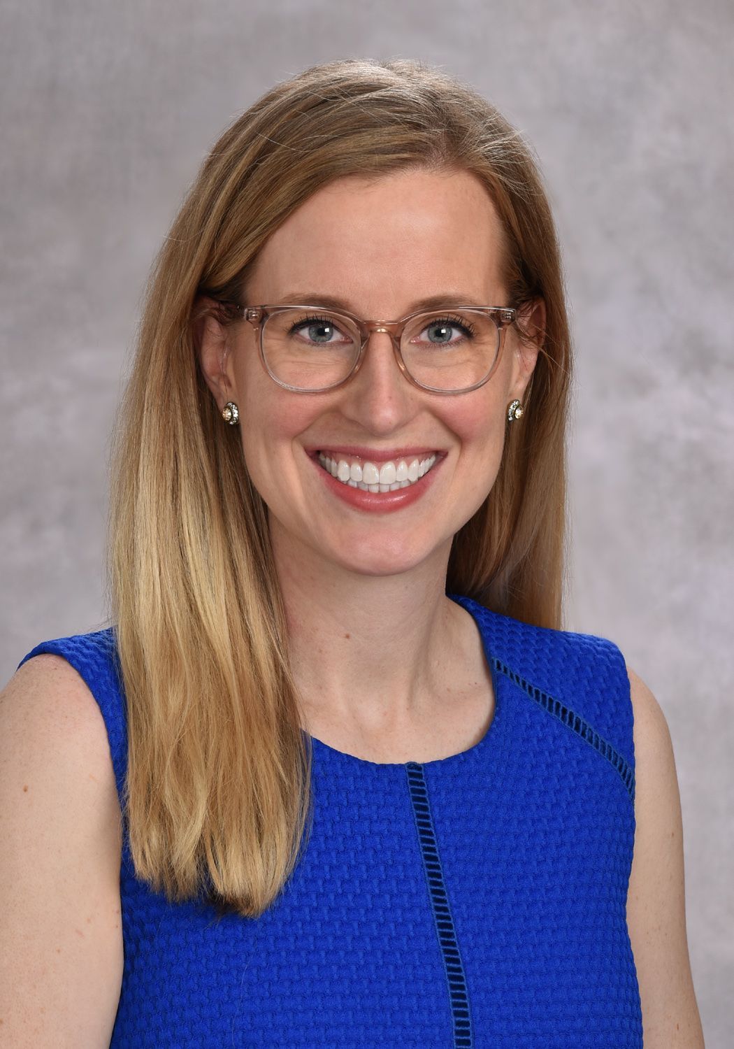
Meanie Gropler, MD
Job title
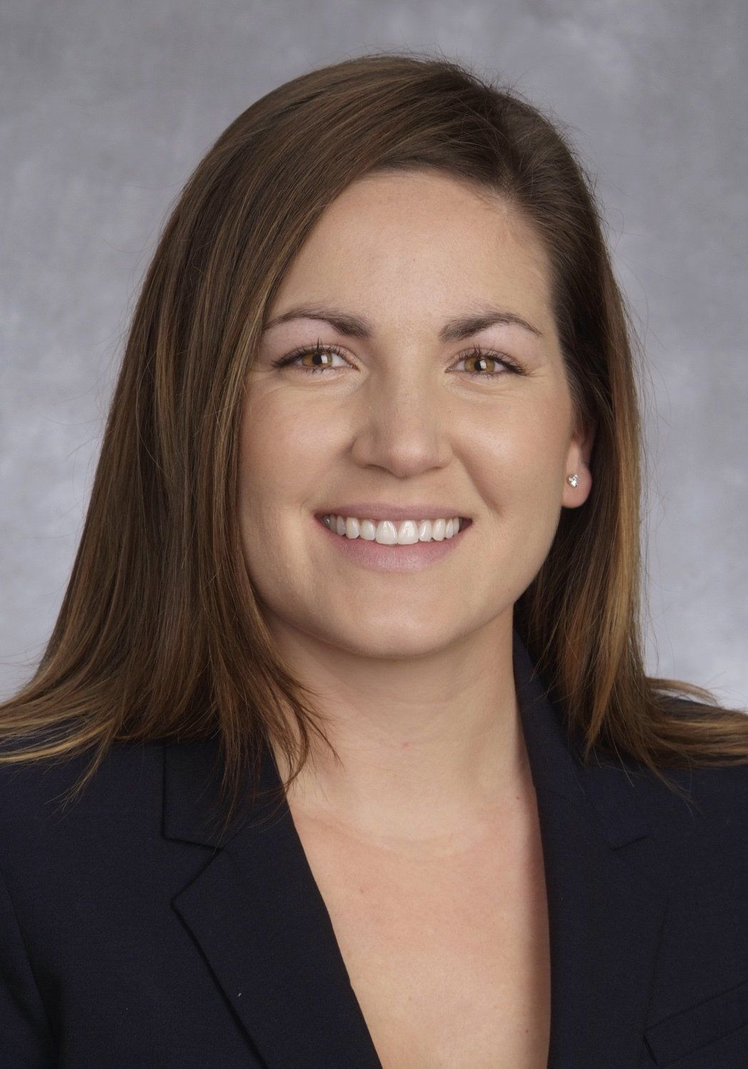
Ericka Hatfield, DO
Job title
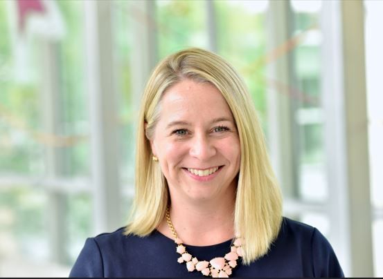
Lisa Howley, MD
Job title